Instrumental diagnostics
Detailed scanning of the whole body with the highest accuracy
JSC "Medicine" conducts a full range of diagnostic studies on modern high-tech equipment and invites patients from 18 years of age and older for a detailed examination of the body to assess the state of health.
Timely diagnostics allows:
- assess the state and work of a particular organ or system of the body
- prescribe an effective and safe course of treatment
- make an informed decision about surgery
- undergo a preventive examination
- detect a dangerous disease and take measures to eliminate it at the initial stage of the disease, when the probability of recovery is 90-100%.
Please note: instrumental diagnostics is carried out strictly if there are appropriate indications and is prescribed by the attending physician. Such a measure is necessary to create a safe environment during the examination and to exclude the risk of deterioration in health due to external influences on the body.
The list of instrumental studies in JSC "Medicine" (clinic of academician Roitberg)
The diagnostic center of the clinic conducts the following types of instrumental examinations:

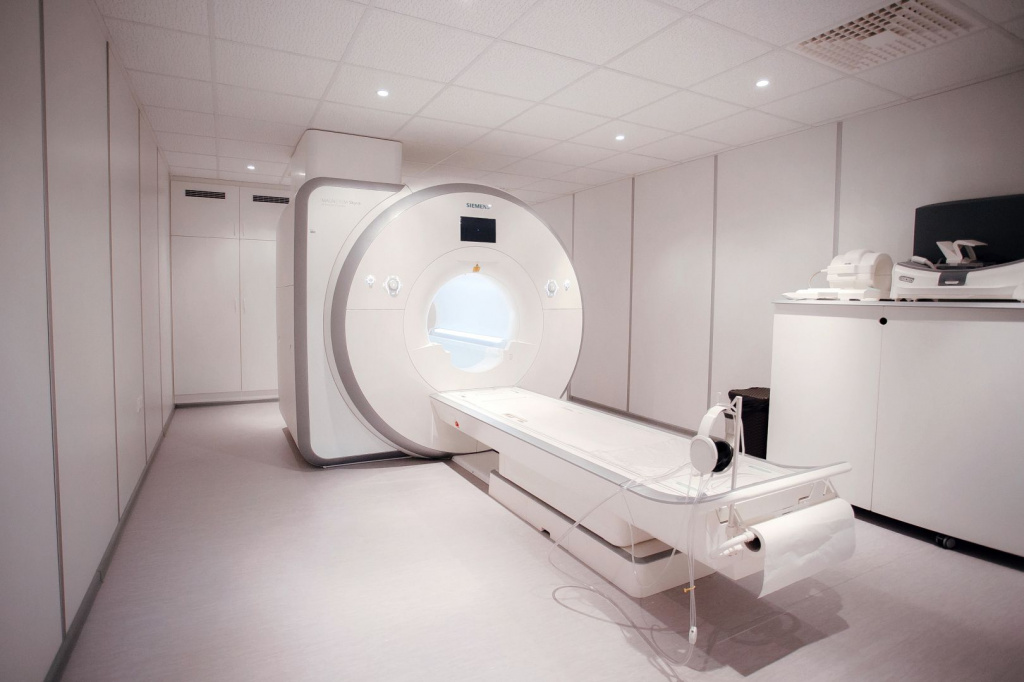
Magnetic resonance imaging is a non-invasive, accurate and safest diagnostic method that allows you to identify a number of diseases and pathologies at an early stage of formation and development. It is used for making an accurate diagnosis, monitoring the treatment process and recovering from surgery. The diagnostic principle is based on the use of high-power magnetic fields without ionizing radiation, which allows you to get a clear image of the internal organs. The examined organ is visualized on the tomograph screen as clearly as possible, since the resolution of MRI is extremely high. It is possible to carry out tomography with contrast - the introduction of a dye into the blood vessels. This makes it possible to clarify the state of blood vessels and tissues of internal organs, to identify areas of stagnation of blood and places of its accumulation due to pathological proliferation of tissues, and also to assess the performance of various body systems.
- the presence or symptoms of oncological formations or benign tumor processes
- edema and metastasis
- joint damage, sprains
- vascular pathology - thrombosis, aneurysm, etc.
- presence of stones in the urinary tract
- pathology of the nervous system
- hernial formations in the spine, etc.
- neuroimaging of cerebral vessels
- The ability to obtain the maximum number of high-resolution images in 20 minutes of scanning;
- The slice thickness during scanning is up to 0.5 mm, which makes it possible to form 3D images with their subsequent reconstruction in any plane without loss of quality. This is necessary to determine the pathology in the early stages and allows you to identify even the smallest changes in organs and tissues.
- The minimum level of acoustic noise.
- Unique endorectal coil for examining the prostate, cervix and rectum.
- The wide diameter of the tunnel is 70 cm, which allows examining patients with a large weight (up to 250 kg) and claustrophobia.
- Cylindrical field of view for more accurate and reliable display of all anatomical details.
- Short terms of examination and obtaining results within 90 minutes from the moment of diagnosis;
- A guarantee of the accuracy and reliability of the results, which do not need to be checked and refined;
- The possibility of a thorough study of complex diagnostic cases;
- Safety and comfort (can be carried out under anesthesia in a state of sleep, the clinic has the necessary equipment for this);
- No restrictions on the number of studies;
- Approved for use in pregnant women and young children.
The results of MRI diagnostics are provided in printed form along with three-dimensional images and on electronic media. The number of images obtained during the study varies from 300 to 1000, depending on the scanning area and tasks.
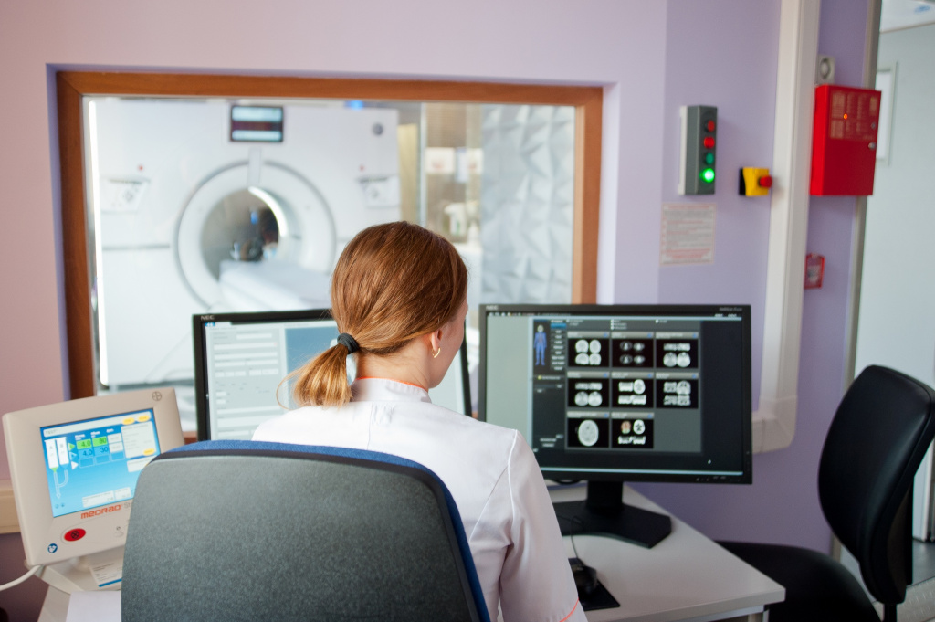
Computed tomography is a popular imaging technique used to obtain cross-sectional scans of any anatomical region of the human body. An important point: the resulting "picture" has a three-dimensional format, which makes it possible to comprehensively assess the state of an organ or area of the body, as well as to obtain sections of internal organs to clarify their condition and performance.
- diseases of the ENT organs
- pathology of the spine, brain or spinal cord
- reproductive system diseases
- disturbances in the work of the heart and lungs
- symptoms of diseases of the abdominal cavity and small pelvis
- pathologies and injuries of the upper and lower extremities.
Diagnosis by computed tomography is carried out strictly in the presence of indications in the direction of the attending physician. CT is not done during pregnancy or for symptoms of diabetes, thyroid disease, or kidney failure.
In the clinic "Medicine", CT of internal organs is carried out on computed tomographs Siemens Definition 64 (128 slices) and Revolution CT GE Healthcare (512 slices).
The Siemens Definition 64 computer tomograph (128 slices) doubles the speed of information collection at low radiation exposure, and GE Healthcare's Revolution CT (512 slices) scans the heart and blood vessels in just 0.2 seconds. This is one complete revolution around the heart. The images are of the highest quality regardless of the heart rate.
- detect pathology at the initial stage;
- get a conclusion within two hours from the moment of examination;
- diagnose a patient of any age;
- evaluate the high speed and accuracy of scanning, as well as detect formations up to 3 mm in size;
- eliminate harm to the patient's health due to low radiation exposure
The equipment of the JSC "Medicine" clinic allows examining patients weighing up to 220 kg.
- Minimum scan duration;
- The ability to obtain images of an organ with a cut of up to 1 mm without additional exposure of the patient to the equipment;
- High resolution images that do not need high detail and allow you to see a high-quality image of internal organs and their structure;
- Acceptable prices.
There are several types of SPECT that are used depending on the purpose of the examination and the preliminary diagnosis:
- osteoscintigraphy allows you to clarify the state of the musculoskeletal system;
- myocardial scintigraphy is aimed at examining the state and functioning of the heart, identifying violations in the structure and functioning of the heart muscle, chambers, etc.;
- scintigraphy of the thyroid gland allows you to identify pathological changes and formations in the structure of the organ, to clarify the presence of inactive nodes, to detect a tumor process, etc.;
- kidney scintigraphy shows the presence of stones, the consequences of trauma, cystic and tumor processes, foci of inflammation and infections, and also allows you to clarify the functioning of organs, parameters of blood flow and urination .;
- brain scintigraphy may indicate pathological disturbances in the blood supply to the brain, disturbances in blood flow in epilepsy and stroke, mental disorders and other diseases, the consequences of injuries and concussions;
- liver scintigraphy indicates inflammatory processes in the parenchyma, vessels and connective tissue, helps to assess the shape and size of the organ, to identify the presence of neoplasms and pathological deformities;
- lung scintigraphy is performed in a perfusion or ventilation format, it can detect blood clots, embolisms, and blood flow disorders;
- Scintigraphy of the parathyroid glands allows you to detect neoplasms, inflammatory processes and vascular pathologies, helps to clarify the parameters of the functioning of organs and identify possible violations of blood supply.
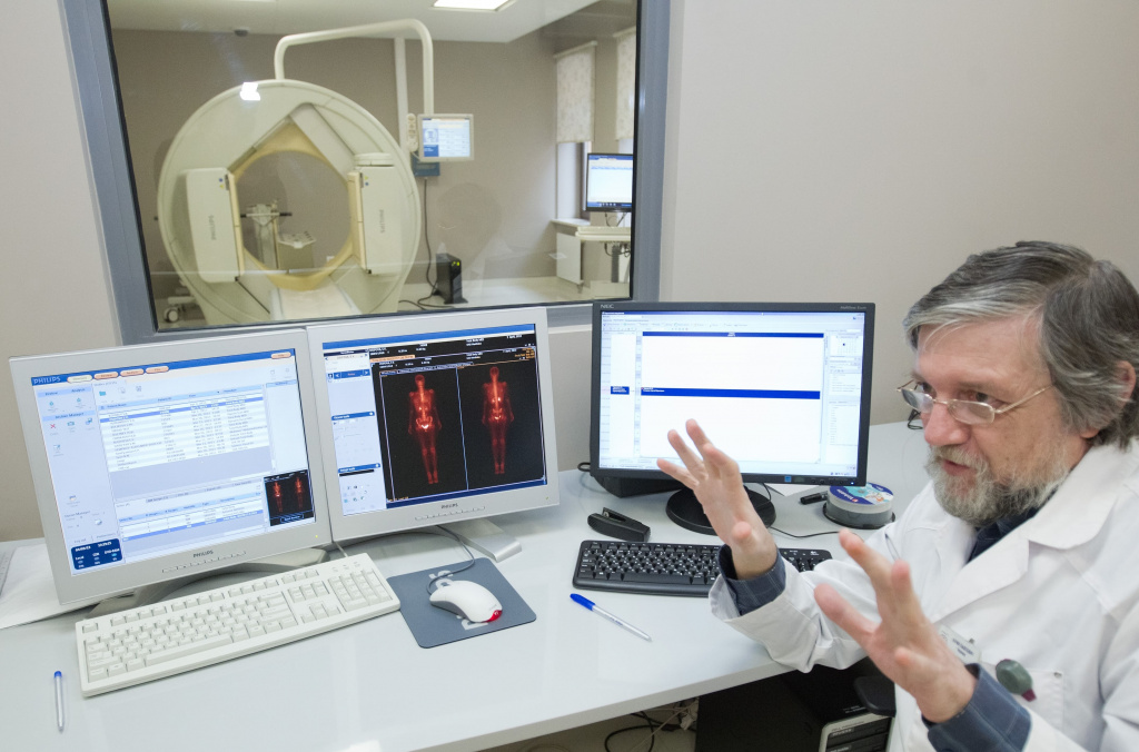
The Medicina Clinic uses the BrightView Philips high-intelligence gamma camera to perform single-photon emission computed tomography (SPECT), which produces high-image images at low radiation exposure. For the study, a radiopharmaceutical (isotope technetium-99) is used, which has a short half-life (only 6 hours) and has a low energy of gamma radiation. It is injected intravenously and accumulates in the organ that needs to be examined. Due to this, the detected foci of the disease can be visualized with high accuracy, which guarantees the correct diagnosis and detection of the disease at an early stage of development.
- The radiation exposure as a result of the conducted study corresponds to an X-ray picture;
- Obtaining an image of planar sections of the organs under study with subsequent reconstruction of their three-dimensional image;
- The ability to select the type of image - a three-dimensional image or a cross-section;
- Detection of the exact localization of a tumor or inflammatory process;
- Visualization of the area of placement of capsules in brachytherapy.
SPECT is used to detect oncological diseases, in order to identify even the smallest pathological focus, regardless of its location, as well as to detect functional changes in the kidneys, thyroid gland, heart, lungs, etc.
Positron emission tomography, combined with computed tomography, is the most informative diagnostic method today, which allows you to most accurately identify the smallest tumor process in the body and see the degree of metastasis of tumor cells. PET / CT is indispensable for assessing the effectiveness of the treatment carried out in the study of the dynamics of a histologically confirmed tumor process, after chemotherapy, radiation therapy and surgical treatment.
For examination, glucose is injected into the body intravenously, combined with a radioisotope, most often "Fluorine-18". Then, within 90 minutes, it spreads throughout the body and accumulates to a greater extent in unhealthy, pathologically active cells. Next, a scan is carried out on a tomograph. Due to radioactive fluorine, glowing points are displayed on the screen - foci in which glucose accumulates. These are diseased cells.
- oncological processes in the lungs, liver, intestines, esophagus, on the skin or on the surface of the larynx, on the lips or tongue;
- sarcoma;
- tumor processes in the uterus, appendages, organs of the reproductive system in men and women;
- suspicion of melanoma or lymphoma;
- epilepsy;
- stroke;
- Alzheimer's disease;
- ischemia of the heart;
- Parkinson's disease.
The Medicina clinic uses 2 modern high-tech PET / CT scanners for PET / CT - Philips Gemini TF and Siemens Biograph MCT. All devices have an increased level of radiation protection and allow quick scanning.
- Fast scanning with the highest image quality due to the width of the detector (16 cm compared to 8 cm in conventional tomographs). At the same time, the image capture with one revolution of the tube is doubled, and the scanning time is reduced to 18 minutes (instead of 30 minutes on the previous generation scanners).
- Low radiation exposure that is comparable to conventional CT examination.
- Fast and accurate finding of the area of tumor localization and spread of metastases.
In JSC "Medicine" (clinic of Academician Roitberg) you can perform PET / CT using the compulsory medical insurance system.
List of documents required for PET / CT examination under compulsory medical insurance:
- • passport or other document proving your identity;
- • SNILS;
- • policy of compulsory medical insurance (compulsory medical insurance);
- • direction (form 057 / y);
- • the results of the previous survey for the relevant profile.
- One of the best diagnostic bases in Moscow, equipped with the most modern equipment.
- High diagnostic accuracy and low radiation exposure.
- All studies are carried out 7 days a week from 8.30 to 21.00. Magnetic resonance imaging (MRI) is available 24 hours a day.
- PET / CT under the compulsory medical insurance policy for residents of Moscow, the Moscow region and other regions of the Russian Federation
- Interpretation and issuance of conclusions is carried out within an hour after the research.
- The research results can be obtained on CD-ROM, by e-mail, as well as in the personal account on the clinic's website and in the application.
- Recording is possible on the day of contact or in the coming days after the recording.
- The study is carried out at a strictly appointed time.
- You can sign up for a study by phone, at the clinic administrator, through your personal account on the website and in the mobile application.

