Volumetric formations of the kidneys
Patient L., 71 years old.
I went to a urologist for an annual medical examination.
Medical history.
History of repeated exacerbations of cystitis. She underwent treatment in the clinic of JSC "Medicine" for an exacerbation of cystitis with a positive effect. At the next dispensary examination - general urine tests, urine culture, ultrasound of the genitourinary system. An ultrasound scan revealed a 17 × 12 mm left kidney cyst?
When conducting a survey.
Ultrasound of the abdominal cavity. Ultrasound signs of a cyst of the right lobe of the liver, deformity, thickening of the walls and the presence of gallbladder polyps, diffuse changes in the pancreas.
CT scan of the chest. Focal, infiltrative changes in the lungs were not identified. Diffuse pneumosclerosis.
Examination by a physician. Stage II hypertension. Stage I dyscirculatory encephalopathy with predominantly circulatory insufficiency in the system of vertebrobasilar arteries. Cephalgic syndrome. Widespread osteochondrosis of the spine.
Laboratory examination. Clinical analyzes without pathology.
ECG. Sinus rhythm. Normal position of the electrical axis of the heart. Signs of early ventricular repolarization.
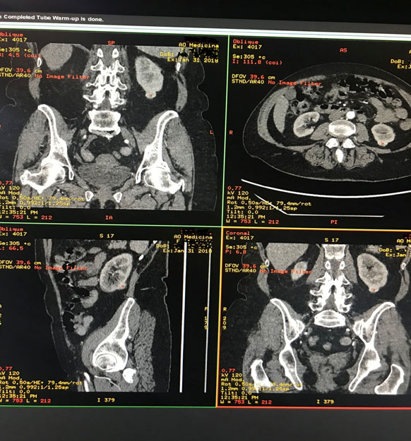
Multispiral computed tomography (MSCT) of the genitourinary system with intravenous contrast. There is a subcapsular formation in the lower pole of the left kidney along the posterior surface (19 × 14 × 16 mm, density 34 HU), in the arterial phase it accumulates contrast up to 134 HU, more along the periphery, in the venous phase - 101 HU, in the delayed phase - 67 HU ... The surrounding fiber is not changed.
The calyx-pelvic system and ureters were not dilated, no radiopaque calculi were found. The renal arteries and veins are not changed.
Данные объективного осмотра.
The condition is satisfactory. Body temperature 36.5 ° C. The skin is of normal color, moderate moisture, clean. Peripheral lymph nodes are not enlarged. No edema. The respiratory rate is 16 per minute. Auscultatory vesicular breathing, wheezing is not heard. Heart sounds are clear, the rhythm is correct, noises are not heard. Heart rate 75 per minute. Blood pressure is 135/75 mm Hg. Art. The tongue is moderately coated with a white coating, moist. The abdomen is moderately distended, soft on palpation, painless in all parts. The area of the kidneys is not changed, painless on palpation, a negative tapping symptom on both sides. Urination is free, painless, no dysuria.
Preliminary diagnosis: volumetric formation of the left kidney.
Performed laparoscopic resection of the mass of the left kidney without thermal ischemia. Blood loss 50 ml. The duration of the operation is 55 minutes.
Histological conclusion. The histological structure corresponds to renal cell carcinoma. The tumor does not grow outside the kidney capsule. The drug is removed within healthy tissues.
Final diagnosis: left kidney cancer T1aN0M0.
Control examination. CT of the genitourinary system after 3 months (condition after resection of the formation of the left kidney): CT signs of continued growth were not detected.
Thus, on the basis of this clinical case, the need for prophylactic medical examination is confirmed to detect cancer at an early stage and timely high-tech endoscopic treatment without further consequences affecting the patient's quality of life and the work of his organs.
For reference.
1. Epidemiology. Kidney cancer is one of the most common cancers in urology. The highest share in the annual growth of oncological diseases is 30%.
2. Diagnostics. The disease (in about 40% of cases) is detected by chance, during a dispensary examination. At the moment when the first symptoms appear, we can talk about an advanced stage. The most accessible method for detecting kidney formations is an ultrasound scan, which is recommended at least 1 time per year. Contrast-enhanced renal computed tomography (the gold standard of renal examination) is used to confirm a suspected ultrasound diagnosis. Unfortunately, at the time of initial diagnosis, distant metastases can be detected in 30% of patients. In the case of controversial data obtained with MSCT, additional information for making a decision on the method of treatment and observation can be obtained by magnetic resonance imaging of the kidneys.
3. Treatment. Depending on the stage of the identified process, various types of surgical treatment can be used. The best results are achieved by laparoscopic (through puncture) operations. At the stage of the disease T1a-b, an organ-preserving operation (resection) is performed, at T2-T4 - laparoscopic radical removal of the kidney with the surrounding structures (nephrectomy). With the timely removal of the tumor focus, a complete cure can be achieved.
4. If a kidney formation is detected, you can receive highly qualified assistance in the clinic of JSC "Medicine", the first in Russia accredited by the Joint Commission International (JCI) in 2011. High-quality staff, comfortable conditions of stay, expert-class equipment, the shortest recovery time are waiting for you.

