


Dental X-ray
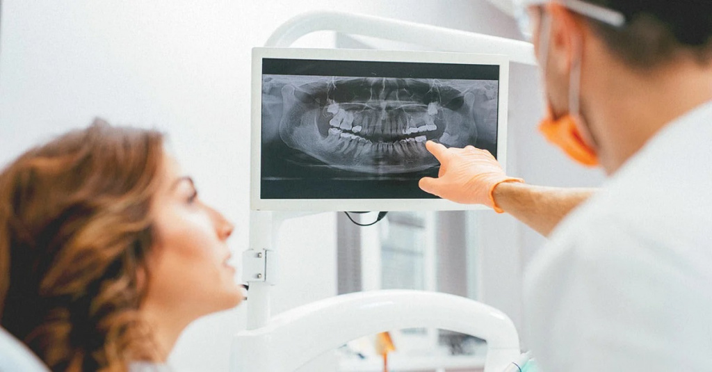 One of the traditional and very popular diagnostic procedures in the dental industry is the radiological examination. What does a dental X-ray show? Dental X-rays can determine the extent of hard tissue damage, identify hidden caries, assess the quality of previous dental treatment and promptly diagnose periodontal inflammation. X-rays can clearly demonstrate not only the condition of the bone tissues, but also identify many negative changes. This is essential for making an accurate diagnosis and determining the appropriate treatment regimen.
Whereas X-ray machines used to be outdated, they have now been replaced by more modern radiovisiographs, which offer a number of advantages:
- Minimal radiation exposure.
- Quick examination time.
- The results are available on the day of the examination, which is very important for a timely diagnosis.
In JSC "Medicina" Clinic you can undergo highly accurate dental diagnostics with the use of a modern X-ray machine, which guarantees safety of your body. It is possible to make an appointment for the procedure by phone or using a special online form on our website.
Are dental X-rays harmful?
Many patients at dental clinics wonder whether dental X-rays are bad for their health. According to the Sanitary and Epidemiological Norms, the permissible level of gamma rays is 1000 microsieverts (µSv) per year. A special feature of modern radiological examinations is the fact that the exposure time of the machine is kept to a minimum, so the radiation dose is negligible.
In terms of specific numbers, one procedure using a digital radiovisiograph is equivalent to 2 microsieverts. As for the film study, the dose is 10 microsieverts.
When answering the question of how harmful dental X-rays are, it should be noted that the frequency of the examination is determined by the attending physician. On average, you can take about 100 shots per calendar year. Obviously, even in a complicated clinical case, so many radiological examinations will not be necessary.
Indications for dental X-rays
A routine checkup at your dentist can only detect external damage. In cases where you want to see what is going on in the tissue, you have to resort to hardware-based methods of examination.
So why are dental X-rays needed? They can be used to diagnose the following abnormalities:
- periodontitis;
- abscess;
- parodontitis;
- concealed caries;
- abnormalities of the TMJ;
- cysts and other neoplasms.
The detection of abnormalities is not the only indication for this examination. Dental X-rays are also mandatory in the following cases:
- treatment of dental canals;
- before placing implants;
- after a tooth resection has been carried out;
- dental prosthetics.
Thus, dental X-rays are a common procedure that is prescribed for all types of endodontic and surgical treatment, as well as in the process of diagnosing deep caries and pathologies of the maxillofacial area.
Contraindications
The radiation dose during an X-ray examination is minimal. But this does not mean that this procedure can be carried out without any restrictions. The main contraindications include:
- pregnancy;
- problems with low immunity;
- mental disorders;
- bleeding.
Main types of dental X-rays
X-rays are a fairly broad concept that includes a number of diagnostic manipulations that involve the use of various types of equipment. In dentistry these are widely used:
- extraoral radiography;
- intraoral radiography;
- electro-roentgenography;
- CT;
- orthopantomogram;
- teleroentgenogram;
- X-ray procedure with a contrasting agent.
Extraoral radiography
This type of examination is carried out when an intraoral radiograph is not possible for some reason. This can be a trismus of the jaw or a gag reflex in the patient. The disadvantage of this method is the lack of accuracy in displaying the oral tissues.
Intraoral radiography
This type of examination is carried out using a special film (5 x 6 or 6 x 8 cm), which the patient has to press between the teeth. It is not allowed to move during the procedure and it is necessary to hold your breath. Otherwise the picture may not come out clearly. However, this recommendation applies to all types of pictures.
Electro-roentgenography
This examination differs from traditional X-rays in the following ways:
- The image is sharper and more informative, so that even the smallest nuances are visible.
- It is possible to determine the density, condition and volume of the periosteal tissues.
- These scans can be used to diagnose areas of haemorrhage, cysts and other masses more accurately.
CT
This examination is carried out using a special apparatus called a CT scanner. It is based on a 3D digital image (3D visualisation effect). The main advantage of this method is its high informative quality, because the image reproduces the projection of the jaw in a three-dimensional version. The specialist can easily examine the condition of the bone tissue and assess the degree of disease and pathological processes. The impact of radiation is low because of the short duration of the procedure.
Orthopantomogram
This is a type of examination that is in high demand. With this technique, it is possible to image absolutely all the jaws and teeth.
Teleroentgenogram
This type of diagnosis allows not only the jaw structure to be examined, but also a facial and a profile picture of the entire skull. This is one of the most informative diagnostic procedures in dentistry. This procedure is performed as a must before the placement of braces and it is also commonly used before upcoming surgeries as a preparatory measure.
Use of a contrast agent
The use of an X-ray with a contrast agent (sialography) is indicated if the condition of the salivary gland ducts needs to be assessed. They are filled with iodine-containing solutions before the procedure. This type of diagnosis can detect a number of abnormalities: salivary stone disease, inflammatory foci and the presence of concrements.
Where can it be done?
If you need to undergo an X-ray examination, you can use the services of JSC "Medicina" Clinic. We have all the necessary equipment to take pictures of high quality and in great detail, safe for your health and the health of your children.
If you have decided to make an appointment with a specialist at JSC "Medicina" (Academician Roytberg Clinic) in the Central Administrative District, please call +7 (495) 845-39-61. We are located in the centre of Moscow at the address: 2nd Tverskoy-Yamskoy Lane, 10. The following metro stations are nearby: "Mayakovskaya", "Belorusskaya", "Novoslobodskaya", "Tverskaya" and "Chekhovskaya".