MRI of cerebral vessels
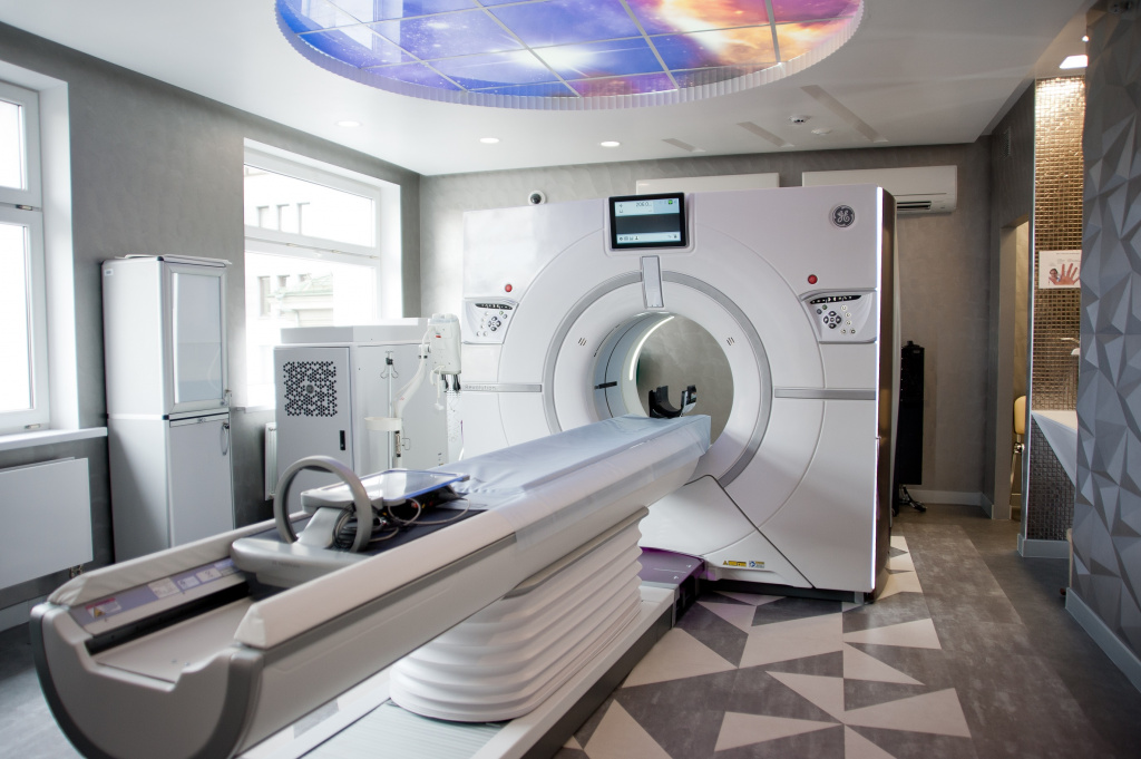
JSC "Medicine" (Academician Roitberg clinic) conducts all possible types of magnetic resonance imaging. To do this, we have installed the latest high-power tomograph (MAGNETOM Skyra power 3 Tesla) and a noise reduction system. These functions ensure maximum accuracy of the study, as well as patient comfort during the procedure. MRI of the brain and blood vessels visualizes them and nearby tissues, providing an opportunity to detect pathological changes even of the smallest size.
Get consultationEquipment
- Up to 250 kg
- Children and adults
- Probably under anesthesia
- With or without contrast
* - The working load capacity of the table is 250 kg, but the decision on the study is made by the doctor, based on the presence of the aperture of the device 70 cm
Its main differences from similar devices of previous generations are as follows:
Maximum clear image
The magnetic field strength of 3 Tesla allows you to explore the deepest layers of tissues and organs without loss of accuracy and create a clear, informative image.
High-precision detailing
64-slice technology is revolutionary today, as it provides high-precision image detail and allows you to register even the smallest signs of pathology, which makes it possible to diagnose oncological diseases at the earliest stages!
Suitable for the youngest patients
The power of our 3 Tesla device allows us to obtain a high-quality image in a shorter time, which facilitates the procedure of MRI of cerebral vessels for children.
No restrictions
Our device is equipped with a table with a high load capacity, thanks to which we can perform MRI of the head of patients with a large weight (up to 250 kg).
Tim 4G Technology
This is a unique development of SIEMENS, which has changed magnetic resonance imaging. Thanks to her, there was no need to change the patient's posture when examining several areas of the body, and high-precision visualization of processes in real time became possible.
The above features make it possible to create as accurately as possible a computer model of brain tissues that displays structural disorders and allows you to make a correct diagnosis based on objective data. This is the most modern equipment for performing magnetic resonance imaging of cerebral vessels. If you need an extremely accurate and objective result, then it will be provided by an examination at JSC "Medicine" (Academician Roitberg clinic).
Indications for MRI of cerebral vessels
It is important to note that magnetic resonance imaging of the brain arteries is often performed simultaneously with brain tomography, since this allows you to get the most complete picture of the disease. But the studies differ somewhat among themselves. So, the brain is examined for various injuries or suspected pathology of the brain substance. Diagnosis of cerebral vessels is carried out if the doctor assumes the presence of vascular diseases: atherosclerosis, diabetic angiopathy, aneurysms and other diseases:
- ischemic disease;
- vascular dementia;
- early manifestations of stroke;
- destabilization of the pituitary gland;
- meningitis;
- suspicious neoplasms;
- blood clots;
- epilepsy;
- headaches;
- Parkinson's or Alzheimer's disease;
- cysts, benign or malignant formations.
The patient may be prescribed a vascular examination, as well as brain arteries for chronic head pain and dizziness, chronic weakness, tinnitus and apathy. As a supplement, tomography is used in the study and treatment of migraines, causeless sudden pressure surges, rehabilitation after a stroke and in the case of neck or skull injuries. Simultaneously with the detection of deviations, their dynamics are evaluated, as well as the functionality of the vessels as a whole. In the case of stroke, tomography allows you to diagnose its form and prescribe the most effective treatment regimen.
Preparation for MRI of cerebral vessels
It is not necessary to prepare specially for a brain vascular tomography, however, on the eve of the procedure, it is necessary to consult a doctor about possible contraindications to the study.


When going for diagnostics, be sure to take with you:
- referral indicating the diagnosis and purpose of the study;
- the results of previous radiation examinations (computed tomography, MRI, radiography, ultrasound);
- medical documentation (expert opinions, extracts from medical history or outpatient card, etc.).
Contraindications for MRI of cerebral vessels
MRI of the vessels of the brain and arteries has the following contraindications:
- the presence of metal stimulators and electronic devices in the body that cannot be removed;
- acute phobias or disorders;
- pregnancy and lactation;
- the inability for one reason or another to maintain a stationary state for a long time;
- the presence of an endoprosthesis.
As you can see, tomography has few contraindications, since it is a safe method of examination. Some restrictions are relative, for example, in the case of claustrophobia. In case of fear of confined spaces before the procedure, the patient is given sedatives that give a calming effect. If it is impossible to conduct an MRI, a CT scan is prescribed to the patient or other alternative diagnostic methods are offered.
Our doctors
Magnetic resonance imaging in JSC "Medicine" is conducted by radiologists with considerable experience, numbering dozens of years of diagnostic practice. These are competent specialists with an excellent reputation, members of the European Association of Radiologists


