SPECT (Scintigraphy)
Single-photon emission computed tomography (SPECT) is a modern method of tomographic investigation including administration of radiopharmaceuticals for visualization of their accumulation in the investigated area.
The method is sufficiently information-rich and at the same time not traumatizing and painless for the patient.
In contrast to traditional scintigraphy, gamma-radiation in SPECT is recorded from different sides. This makes it possible to obtain 3D images of investigated tissues and assess the degree of their involvement in the pathologic process due to their ability to accumulate radioisotopes. The more intensive gamma-radiation in the diagnosed region is revealed, the more it is involved in the pathologic process.
The images obtained during a SPECT scan are called scintigrams. They are processed with the help of special software and arranged according to the algorithm providing for the comprehensive picture of the pathologic process.
The main benefit of the SPECT method consists in the fact that it allows to record initial pathologic changes and the smallest tumor foci, and, therefore, it is irreplaceable for early diagnostics.
Scintigraphy allows to scan all bones in the body rapidly and at minimum costs. But scintigraphy also has a shortcoming: it is insufficient for the final diagnosis. Even if any change in the bones is present, an additional check is required to elucidate a cause. The final diagnosis is made after an X-ray, a CT scan, a biopsy, or an MRI scan, taking into account the signs and symptoms reported by the patient.
Indications for Scintigraphy
Oncology is the main area where the SPECT method is used. As it allows to reveal the smallest foci of neoplastic tissue changes, which are indistinguishable by other diagnostic methods, it is often prescribed in case of disputable and doubtful diagnostic results. Additionally, SPECT is widely used in cardiology; it allows to assess the condition of the myocardial tissue, its supply with oxygen and the level of metabolism.
Basic indications for scintigraphy are as follows:
- diagnosing a primary tumor and metastases of different localization;
- diagnosing bone metastases including remote ones;
- diagnosing cardiovascular pathologies;
- investigation of the state of the cerebral circulatory system.
Contraindications
Pregnant (at any term) or breast-feeding women may not undergo the SPECT procedure. Besides that, osteoscintigraphy is contraindicated in cases of severe immunodeficiency conditions. General anaesthesia is required for diagnostics of small children because the patient should remain motionless during the procedure. Therefore, it is advisable to use scintigraphy only in those cases when other methods can not provide the objective picture of the pathologic process.
What Happens During the Scintigraphy Procedure?
Scintigraphy does not require any complex preparation of the patient. A radiopharmaceutical substance is administered to the patient before starting the scintigraphy; then the patient has to wait for a while for the substance to get uniformly distributed through the body. The waiting time is from one to three hours depending on the investigated region. The bladder should be emptied just before scanning.
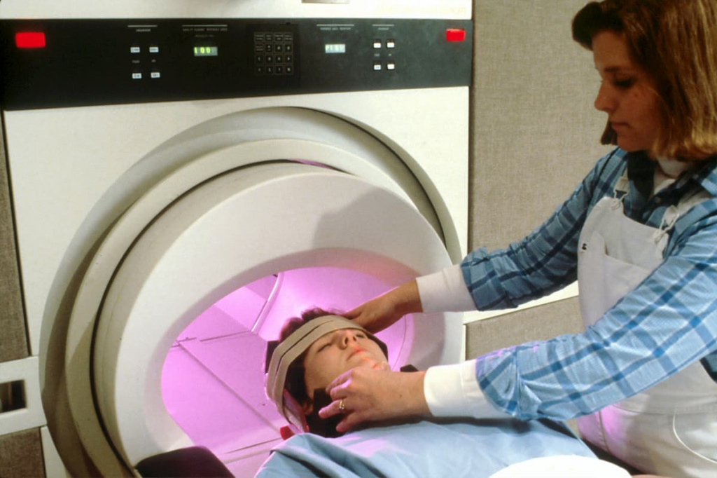
Scintigraphy is performed in two special gamma-chambers which scan the investigated region and the whole body. The procedure takes on average from 20 to 40 minutes. As scintigraphy includes the use of pharmaceuticals, after its completion the patient should increase liquid intake to facilitate the fastest possible excretion of the medicaments.
Diagnostics
Scans are processed by special software. The results are interpreted by a physician. All scintigrams are recorded on electronic media. As a rule, you can get a case report and have a detailed consultation with the doctor in one day.
Advantages of Having a SPECT Procedure Done at JSC «Medicine» (clinic of academician Roytberg)
Cutting-edge equipment at JSC «Medicine» (clinic of academician Roytberg) allows not only to reveal pathologic zones but also to investigate biologic processes in them (pathologic hypervascularization, apoptosis levels, changes in cell replication, involvement of surrounding tissues, etc.). Thanks to high spatial resolution, our scanners can detect the smallest mass lesions (of less than 1 mm) and provide the detailed structured information on the pathologic zones.
Learn everything about your health at JSC «Medicine» (clinic of academician Roytberg)!
Prices
We are trying to offer attractive and competitive prices. The price may vary depending on the type of the procedure. In any case, our prices are affordable for most patients of the Clinic.
Doctors


