MRI of the joints

Magnetic resonance imaging of joints is used mainly in traumatology and neurology. This diagnostic method allows you to determine pathological changes in the soft tissues of the joint: ligaments, cartilage fibers, capsule, meniscus. MRI is sensitive to degenerative and destructive processes inside the articular parts.
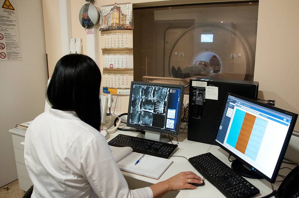
The power of the MRI apparatus in JSC "Medicina" Clinic (Professor Roytberg Clinic) is 3 Tesla. The research is conducted around the clock.
Possibilities of MRI of joints
The magnetic resonance imaging technique is popular and in demand among specialists and patients. The absence of radiation exposure and the high information content of the study allow for diagnostics with the required frequency and without fear for health.
MRI reveals structural features, structure of joint tissues, signs of destructive changes, inflammatory processes and the presence of neoplasms of various sizes and localization. Pictures are taken in several planes at once. To improve the diagnostic capabilities of MRI, contrast enhancement is performed. The study in this case takes place on an empty stomach.
With the help of MRI, it is possible to study all soft tissues located near the joints, as well as bone formations. Sometimes this helps to detect pathologies that affect large areas and involve both bones and articular surfaces.
Indications for MRI of joints:
- inflammatory and infectious joint lesions;
- destructive changes in the vertebrae;
- damage to connective tissue and joints (lupus erythematosus, gout);
- sports or domestic injuries;
- pain, limitation in joint mobility;
- swelling of the soft tissues of the joint;
- the presence of bloody purulent contents in the joint;
- diagnostics of metastases, neoplasms;
- preparation for the operation;
- pathological mobility or joint instability;
- violation of innervation and blood flow;
- dislocation of the joints.
Contraindications to MRI of joints:
- pregnancy, breastfeeding of a newborn baby, kidney disease and diabetes mellitus (with contrast MRI);
- fear of confined spaces, narrow rooms;
- intolerance to contrast media;
- metal objects implanted into the patient's body: insulin pump, pacemaker, etc.;
- mental disorders.
Contraindications are considered on an individual basis. The doctor makes a decision on the need for an MRI, but only after a survey, studying all medical records and assessing the degree of possible risk. It is advisable not to carry out MRI without contrast in the first months of pregnancy, since during this period the most important organs are laid and even a slight effect can harm the unborn child.
Doctors


